

Akimoto T. et al., 2026: Low-Intensity Extracorporeal Shock Wave Therapy Restores Passive Urethral Closure Mechanism in Vaginal Distension Rat Model.
Akimoto T, Ashikari A, Kurobe M, Takamatsu G, Akiyama Y, Kume H, Miyazato M.
Int Urogynecol J. 2026 May 16. doi: 10.1007/s00192-026-06703-7
Abstract
Introduction and hypothesis: Low-intensity extracorporeal shock wave therapy (LiESWT) is a promising noninvasive therapy for stress urinary incontinence (SUI). We assessed whether LiESWT restores urethral function in a vaginal distension (VD)-induced rat model of SUI, focusing on pelvic floor tissue remodeling and endoplasmic reticulum stress pathway as potential mechanisms.
Methods: Eighty-one female Sprague-Dawley rats underwent functional and tissue analyses. Sneezing was induced to assess urethral pressure responses. Leak point pressure (LPP) was measured with or without hypogastric and pudendal nerve transections. Tissue remodeling and molecular pathways were assessed using histological and molecular expression analyses.
Results: In VD rats, baseline urethral pressure (9.9 ± 4.3 vs 17.6 ± 3.1 cmH2O) and LPP (27.4 ± 4.7 vs 37.0 ± 7.0 cmH2O) were lower than in normal rats (both p < 0.05). LiESWT restored the baseline urethral pressure (16.3 ± 4.7 cmH2O) and LPP (37.5 ± 4.9 cmH2O) in VD rats (p < 0.05). Notably, LPP enhancement persisted after bilateral hypogastric and pudendal nerve transections. Additionally, LiESWT tended to reduce VD-induced vaginal smooth muscle fibrosis histologically, whereas collagen type I alpha and elastin mRNA expression remained elevated in both groups. Western blotting revealed no sustained activation of protein kinase R-like endoplasmic reticulum kinase (PERK) at 96 h after the final LiESWT.
Conclusions: LiESWT enhanced urethral function in the VD-induced SUI model by potentially facilitating pelvic floor tissue remodeling, thereby restoring passive urethral closure. The absence of sustained PERK activation in the late phase indicates the safety of LiESWT for SUI.
Comment Jens Rassweiler
Objective
The authors assessed whether LiESWT restores urethral function in a vaginal distension (VD)-induced rat model of SUI, focusing on pelvic floor tissue remodeling and endoplasmic reticulum stress pathway as potential mechanisms.
Study design
81 female Sprague–Dawley rats underwent functional and tissue analyses. To address the objective question, the authors utilized the vaginal distension (VD) model, which simulates the mechanical stress and ischemia experienced by vaginal tissues during pregnancy and vaginal delivery and is widely used to study SUI.
Sneezing was induced to assess urethral pressure responses. Leak point pressure (LPP) was measured with or without hypogastric and pudendal nerve transections. Tissue remodeling and molecular pathways were assessed using histological and molecular expression analyses.
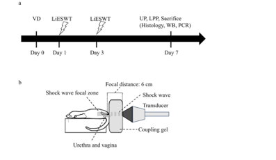
Under 2% isoflurane anesthesia, LiESWT (0.12 mJ/mm2, 2 Hz, 300 shocks/session) was delivered transperineally using an ED-1000® device (Medispec, Yehud, Israel). The transducer was adjusted to an optimal focal depth to target the mid-urethral region. Treatment was performed 24 and 72 h post-VD (Fig. 1a).
Results
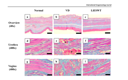
In VD rats, baseline urethral pressure (9.9 ± 4.3 vs 17.6 ± 3.1 cmH2O) and LPP (27.4 ± 4.7 vs 37.0 ± 7.0 cmH2O) were lower than in normal rats (both p < 0.05). LiESWT restored the baseline urethral pressure (16.3 ± 4.7 cmH2O) and LPP (37.5 ± 4.9 cmH2O) in VD rats (p < 0.05). Notably, LPP enhancement persisted after bilateral hypogastric and pudendal nerve transections. Additionally, LiESWT tended to reduce VD-induced vaginal smooth muscle fibrosis histologically, whereas collagen type I alpha and elastin mRNA expression remained elevated in both groups. Western blotting revealed no sustained activation of protein kinase R like endoplasmic reticulum kinase (PERK) at 96 h after the final LiESWT.
Discussion
This study demonstrated that LiESWT restores the passive urethral closure mechanism in a VD rat model of SUI. Thus, it confirms the results of Tom Lue`s groupfrom the year 2024.
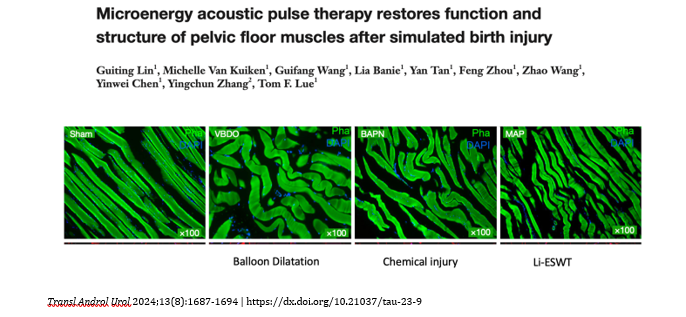
Unfortunately, the authors do not discuss this paper, anyway based on these findings first clinical trials are on its way. The similarity of the findings is striking and may indicate a stimulation of stem cells as main mechanisms resulting in anatomical restoration of the sphinteric muscle.
Conclusion
In this rat VD model of SUI, LiESWT restored the baselineurethral pressure and LPP to near- normal levels. Histologically, vaginal fibrosis tended to reduce after LiESWT treatment. These functional enhancements persisted after nerve transection, indicating that LiESWT restores passive urethral closure mechanism through effects on the periurethral vaginal tissue. This could be based on stem cell stimulation.
The absence of sustained PERK activation at 96 h after the final LiESWT supports a favorable safety profile. These results support further assessment of LiESWT as a noninvasive treatment for SUI.
Jens Rassweiler


Comments